Basic approach to otoscopy
Otoscopy is the assessment of the ear canal and the tympanic membrane ear using a medical device known as the otoscope. This tool can help diagnose otitis externa and otitis media. Generally in adults, the auricle/pinna is pulled upward and posteriorly to straighten the ear canal. In kids, pull the auricle downward and posteriorly for the most direct view. Once a clear view of the ear canal and the tympanic membrane is obtained, it is time to start assessing the ear.
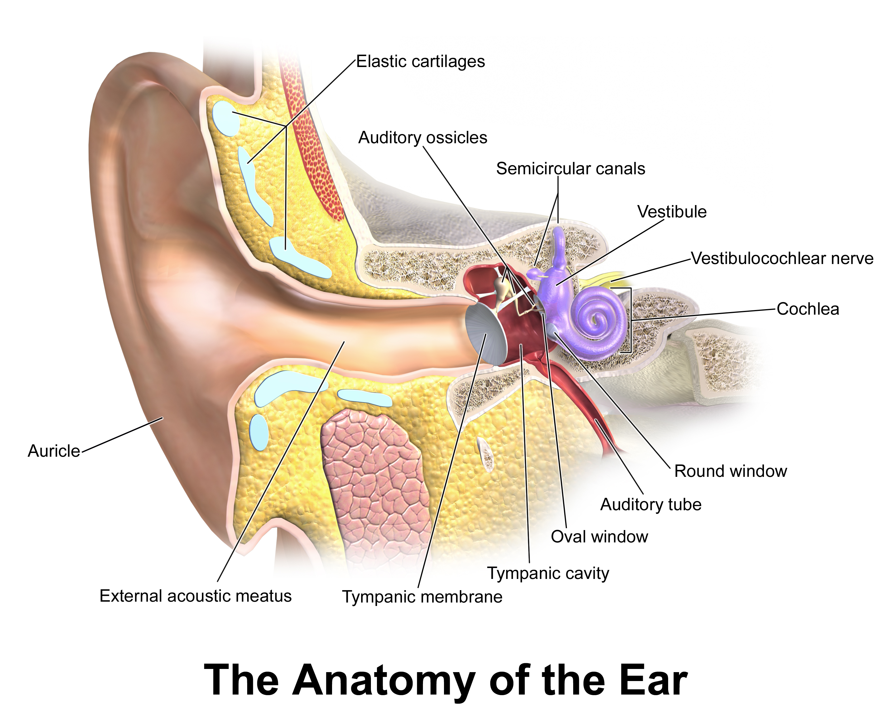
Diagram 1. Quick review of the normal ear anatomy
Sometimes pneumatic otoscopes are used instead of the regular ones. These otoscopes are capable of blowing air on the eardrum. This enables the assessment of another variable, which is mobility. Uninflamed eardrum should be mobile.
Things to evaluate
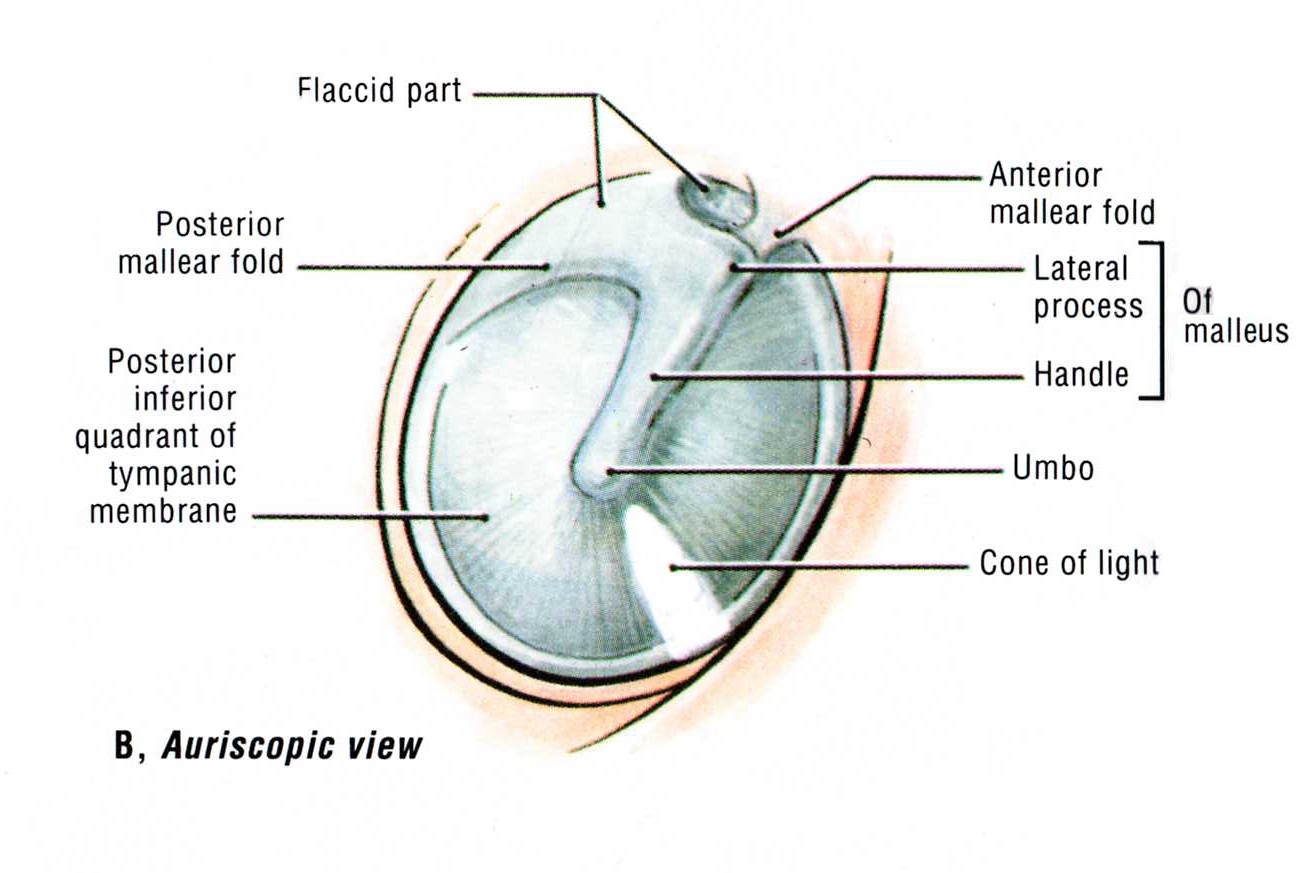
Diagram 2. Parts of the tympanic membrane visible on otoscopy
- Colour of the tympanic membrane
- Can be red, white or yellow
- Pinkish gray is the normal colour
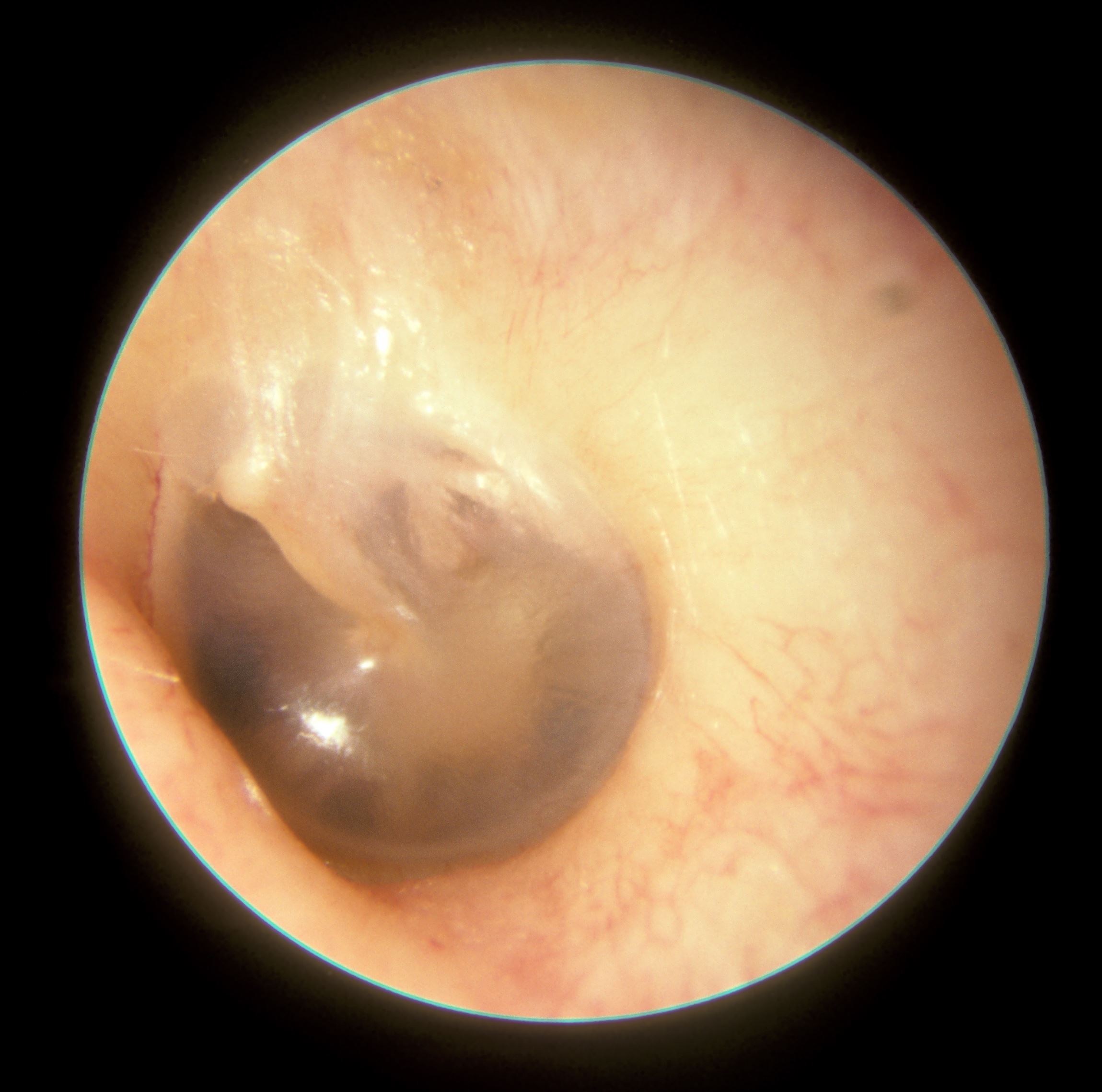
- Translucency of the tympanic membrane
- Opaque or translucent
- Normal should be somewhat translucent
- Position of the tympanic membrane
- Is it bulging, or retracted?
- Identify pars tensa and the cone of light
- Identify the handle of malleus along with the lateral process and ensure proper placement (lateral process will point anteriorly)
- Identify the anterior and posterior folds of pars flaccida
Outside these, comment on the amount of vax, presence of foreign objects, discharge, bleeding and pus.
All information provided on this website is for educational purposes and does not constitute any medical advice. Please speak to you doctor before changing your diet, activity or medications.